3 exercises to prevent low back pain

These 3 exercises are termed ‘the Big 3’ by spinal researcher Dr Stuart Mcgill. The exercises are performed in a neutral back position – a spine sparing position and it helps to build muscular fitness along with stability and control throughout the spinal column. Spinal stability is what creates a strong core.

You do not have to be in pain to perform these exercises. These are actually best used as a preventative method to prevent back pain! Although if you are in pain, they are still a great way to help you get your core switching on again.

Some things to note:

·   The exercises must be performed daily

·   The best time is mid-morning or dinner time. Not upon waking – as this is when the spinal discs are at their fullest and therefore less room to move

·   Reps and sets are important to what you can tolerate. There is no point trying to push through excruciating pain. Find your level and then progress

·   Try and ensure that all 3 exercises are performed in a pain free posture for your spine.

1. Bird dog

Position yourself on all fours. Raise your right hand forward while simultaneously extending your left leg back, until both are parallel. Bring both arm and leg back into the resting position and then repeat with the opposite arm and leg. This is one rep.

Make sure your pelvis has minimal rotation while performing this exercise. The value of this exercise is coordinating upper back, lower back and hip movement all together.

Perform 1 set of 8, followed by another set of 6, with a final set of 4.

2. The side plank

Lie on your side, resting on your bent forearm, with your legs straight out

Lift your hips off the floor and hold for 10 seconds. You should feel your abdominal muscles and obliques (side) working. This exercise also works the quadratus lumborum (QL), a major spine muscle that plays a role in spine flexion and rotation. This exercise gets the  QL and oblique muscles working together in coordination.

Perform 3-4 holds per side of 10 seconds each. Repeat for 3 sets, but with each set, reduce the reps by 1.

3.      The curl up

The Mcgill curl up is not like your traditional curl up. There is no movement from lumbar spine.

Start by lying on the ground, with 1 leg bent up and the other leg straight (the painful side should be the one with the straight leg). Place both hands under the small of your back, this will ensure minimal movement through the lower back.

Lift your head off the ground by a few inches and then hold it there for a count of 10. The goal is to perform this without any movement in the lower back. If you feel your lower back rounding, then your head or shoulders are too high off the ground.

During this, you should feel your abdominal muscles brace – this is the spinal stability

Repeat another 5 times for a total of 6 reps. The recommended volume is 1 set of 6, followed by another set of 4 and the final set of 2. You do not need to hold the curl up for more than 10 seconds, rather to progress you can increase the reps.

Conclusion

I recommend using these exercises every day as the best preventive measure to avoid lower back pain. If you do have back pain, they are also great to perform as they can be done in a pain free position. The key is being consistent, once a day and before any exercise.

Mitchell Roberts – Chiropractor

Disc injuries, what actually happens?

Disc injuries are a very common presentation to the chiropractor. We often hear a patient say they have previously had a ‘slipped disc, which sounds like a very painful injury! The good news is that it is actually not possible for the disc to slip out of place, as it held together through some very strong ligaments to the surrounding vertebra. I have detailed below some of the most common disc injuries. I also discuss a disc bulge, a very common phenomenon that does not always lead to low back pain.


In the lumbar spine, there are many different structures that can be the cause of one’s low back pain. Some of these causes can be from the vertebral joint, muscles, ligaments, nerves and the intervertebral disc. It is very important to ensure a thorough history and orthopaedic/neurological examination can help identify the cause and thus the appropriate treatment and exercises provided.


Anatomy of the intervertebral disc:

The discs are located between the vertebra and act as a shock absorber and also help distribute the load through the spine during day to day movement.

There is a total of 23 discs in the human spine – 6 in the cervical (neck), 12 in the thoracic (mid back) and 5 in the lumbar (low back). The disc is made up of a tough outer layer of cartilage (annular fibrosis) which is weaved together like a basket of 15-25 layers. The inner layer is called the nucleus pulposus, which is a gelatinous centre. (A common analogy is a jam donut. The nucleus is the jam and the outer layer is the dough).

Below, I am going to discuss a disc bulge, herniation and finally an annular tear. These definitions and classifications came about through a meeting of several professions/groups (Fardon, 2001) to clarify and standardise disc injury terminology.

Disc Bulge:

With a bulge, the structure and integrity of the disc is still intact. There is a general enlargement of the disc beyond its natural boundary although the contents of the disc are still contained.


A disc bulge can often cause no pain at all. In fact, a 2015 review by Brinjikji found that in people with no back pain at all, 30% of 20 year olds had a disc bulge, with it increasing to 84% of 80 years olds having a disc bulge. A bulging disc is in fact not a diagnosis, it is more of a descriptive term for the shape of the disc contour.

So why is there such a high percentage of individuals with no back pain having disc bulges? The answer being, what structures does the disc touch, irritate or compress. Often a disc bulge is not the source of the pain, rather an incidental finding on imaging.

Disc Herniation

A herniation is defined as a localised displacement of disc material beyond its limits of the intervertebral disc space. Herniated discs result from damage to the tissue structure, resulting in  leakage of contents. They are classified based upon their shape – either an extrusion or protrusion.

The symptoms of a herniation vary from a minor ache in the lower back and buttock, to a more severe pain that radiates down the leg, pins and needles, numbness and weakness. Often the patient leans to one side – to take pressure off that area of the disc that has herniated.

Please note that in rare circumstances, a herniation may cause a loss of control of bowel and bladder functions, which is a medical emergency.


  • Protrusion – A broader base, defined as being wider that it is tall.


  • Extrusion – has a thin ‘neck’ and the extruded material is longer than it is wide. A sequestration can also occur, if the extruded material breaks off at the neck.

Annular Tear

A tear can result to the outside layer of the disc, which can cause local inflammation/swelling which hence irritates the surrounding spinal nerves, resulting in sciatic pain. There is no herniation of the disc contents. It should be noted that some tears can show up on scans in asymptomatic people and thus can be an older injury that has recovered.


Do Discs heal?

The answer is yes, discs do heal and 60-90% of lumbar disc injuries can be treated with a conservative approach, such as chiropractic treatment. (Chui, et al,. 2015).

Based upon all the different type of injuries that can occur to the discs, it is important to make sure a thorough history and examination is performed. Sometimes an MRI may also be needed to show the extent of the injury and be graded. From then on, we can get a clearer picture of what the recovery time will be like for such an injury.

Mitchell Roberts  – Chiropractor


References:

Brinjikji, P.H. Luetmer, B. Comstock, B.W. Bresnahan, L.E. Chen, R.A. Deyo, S. Halabi, J.A. Turner, A.L. Avins, K. James, J.T. Wald, D.F. Kallmes, J.G. Jarvik. Systematic Literature Review of Imaging Features of Spinal Degeneration in Asymptomatic Populations. American Journal of Neuroradiology Apr 2015, 36 (4) 811-816

Chiu, C. C., Chuang, T. Y., Chang, K. H., Wu, C. H., Lin, P. W., & Hsu, W. Y. (2015). The probability of spontaneous regression of lumbar herniated disc: a systematic review. Clinical rehabilitation, 29(2), 184-195.

Fardon, D. F., & Milette, P. C. (2001). Nomenclature and classification of lumbar disc pathology: recommendations of the combined task forces of the North American Spine Society, American Society of Spine Radiology, and American Society of Neuroradiology. Spine, 26(5), E93-E113.


How to make our spinal discs stronger

It has long been thought that the constant pounding of running can cause degeneration of the lumbar disc, however this theory has recently been debunked. A 2017 study from Deakin University in Victoria has found that people who regularly run or walk have stronger and healthier discs in their spine compared to people who do not exercise.


It was shown that through running or brisk walking, the disc showed improved hydration, increased protein content and growth of the disc. The control group for the study were sedentary, non-active adults – whom showed no change in disc characteristics.

One important factor from this study is that the brisk walking and running groups both showed the same amount of improvement in disc health. So, you don’t have to be a runner to improve the health of your discs, walking is fine as well!


The authors concluded that the response of the disc to running is very similar to the response of muscle in resistance training.


What if I have a disc injury?

If you have a disc injury, movement is good for it! It allows the disc to recover through getting nutrition to the disc to allow for repair. If minimal movement occurs at the disc, healing will be therefore very slow.


Why is this so?

Discs get their nutrition through diffusion from the bony endplate of the vertebra above and below. The way this diffusion works is through movement. The constant loading and unloading of the spine through walking or running allows for the flow of nutrients to the disc. In sedentary individuals, the diffusion of nutrients is poor due to the lack of movement, hence why degeneration and dehydration of the discs is more likely.


So the take home message is to move more to strengthen and improve your back health. It also doesn’t have to be intense running, a brisk walk is also as beneficial. Walking with a disc injury is also an important part of the recovery, as it allows for the disc to receive nutrients to allow for repair to occur.


Belavy, D. L. et al. Running exercise strenghtens the intervertebral disc. Sci. Rep. 7, 45975; doi:10.1038/srep45975 (2017)

Mitchell Roberts – Chiropractor

Is it safe to exercise when you have the cold/flu?

With winter here, it is that time of the year for the cold and flu season. Getting sick and taking time off of training / exercise can be a real setback for anyone who has put in the hard work throughout the year in achieving their fitness goals.

The question often arises, ‘how do I know if training will make me even more sick’? A general rule is the “neck check”. If you have symptoms in the head or throat, it should be ok to do light / moderate intensity exercise. If the symptoms are below the neck, it is best to rest.

Following the neck check rule, you can exercise when suffering from:


  • Runny / stuffy nose
  • Watery eyes
  • Mild headache
  • Mild sore throat

It is important to remember that you need to keep the intensity to moderate. Do not try and attempt a record weight or perform reps to fatigue. Stay hydrated and ensure adequate rest after exercise.


Avoid exercising when suffering from these below the neck symptoms:

  • Chest congestion
  • Nausea
  • Fever/chills
  • Coughing up mucus
  • Joint/muscular aches
  • Diarrhea

The benefit of exercising throughout the winter months – it helps to prevent respiratory infections. A 2012 study from Barret et al, found that moderate aerobic exercise of 30-45 minutes duration can half the risk for respiratory infections. Examples of exercises can be walking, running or cycling. So one of the best preventative techniques for avoiding the cold/flu this winter is to get moving!

  1. Meditation or Exercise for Preventing Acute Respiratory Infection: A Randomized Controlled Trial. Barrett et al. Ann Fam MedJuly/August 2012 10 no. 4 337-346

Mitchell Roberts – Chiropractor
The 3 best exercises to prevent low back pain

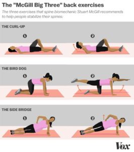
A question I often get asked in clinic is what are the best exercises to perform to prevent low back pain. The below exercises are my ‘go-to’ for prevention of low back pain. They have been termed ‘The McGill big 3’ – named after the researcher Stuart McGill.

Stuart McGill is a spinal biomechanics researcher from The University of Waterloo, Canada. These role of these 3 exercises is to create spinal stability and endurance – which is essential for creating a stable foundation for the lower back.

The exercises aim to tighten up the front and sides of the core, while supporting the spine and remove gravity from the equation. The exercises are of a low intensity and are safe to perform.

Curl up – 3 sets of 10-12 reps

Bird dog – 3 sets of 10-12 reps

Side bridge – 3 sets of 8-10 seconds holds, each side.

If the above rep scheme is too hard, you can always modify it by reducing the number of reps and work your way up to the noted rep scheme.

It is also recommended to do these exercises before training as well, as it has shown to tighten and stiffen the core post performing them.

Perform these exercises once a day, not into pain. If you do have questions, pain or discomfort when performing these exercises, come in and see us and we can either correct your technique or advise you on alternative exercises.


References:

McGill, S.M. (1997) The biomechanics of low back injury: Implications on current practice in industry and the clinic. J. Biomech. 30: 465-475.
McGill, S.M., Low Back Disorders: Evidence based prevention and rehabilitation, Human Kinetics Publishers, Champaign, Illinois, 2002.

Mitchell Roberts – Chiropractor 
My knee/shoulder clicks, does that mean I have arthritis?

I often have patients presenting to me complaining of repetitive pain-free joint clicking (crepitus) of their knee or shoulder. This can often cause anxiety as they associate this clicking with arthritis and joint degeneration. It tends to be more common in the morning and after a period of inactivity, though reduces with movement / once warmed up.

A 2017 study by Robertson et al found that knee crepitus lead to worry, anxiety and eventually fear-avoidance behaviour – meaning they stopped doing their regular exercise because of the sounds.

Joint clicking


Thankfully, joint crepitus is entirely normal. A 1987 study by McCoy et al found that of 250 normal knees studied (no prior injury and pain free), 99% had knee crepitus! 

So what is causing the noise? First off, it is not bone on bone – as this would be very painful and uncomfortable. The noise is fluid moving within the joint and/or tendons/ligaments rubbing and flicking against the bone on the outside.

The role of health practitioners should be to educate patients and eradicate their fear that no damage is being caused by the sounds.

Knee Crepitus = Normal 

The concluding message is that clicking and creaking in joints is very common and is not a sign of joint damage – as long as there is no associated pain or swelling. If you do have any concern, come in and see us at Mona Vale Chiropractic Centre for a thorough assessment.


McCoy G, McCrea JD, Beverland D, Kernohan G, Mollan RB. Vibration arthrography as a diagnostic aid in diseases of the knee. J Bone Joint Surgery (Br) 1987; 69-B, 2: 288-293
Robertson CJ, Hurley M, Jones F. People’s beliefs about the meaning of crepitus in patellofemoral pain and the impact of these beliefs on their behaviour: A qualitative study.Musculoskelet Science and  Practice. 2017 Apr;28:59-64

Mitchell Roberts – Chiropractor


Rigid vs Kinesio Taping

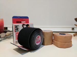
Another common question I get asked in clinic is the difference between the two most used taping methods – rigid and kinesio taping. In short, rigid taping is used for joints such as ankles and shoulders to provide added support, while kinesio tape is used for muscles and joints. They work very differently, which I will explain below.

Here at Mona Vale Chiropractic Centre, we use both forms regularly.

Taping

Rigid Taping:

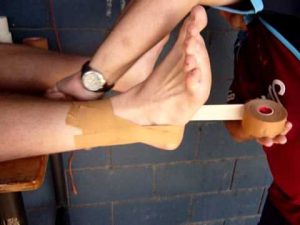
Rigid taping is the most common technique and has been used for a very long time in sport. It is used primarily on joints such as ankles, knees, wrists, elbows and shoulders. The role of the tape is to physically support the joint by limiting movement. The tape is primarily used in returning to sport after a joint sprain, for example an ankle sprain. It provides support and stability that gives the athlete confidence within the joint. It is commonly used in rugby union/league, AFL and netball.

Kinesio Taping:

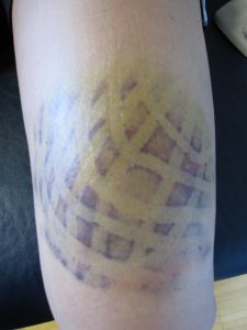
Kinesio taping works entirely different to rigid. The tape is quite stretchy and is applied in a stretched position. Because it is stretched, it bunches once applied. This bunching lifts up the different layers of skin, fascia and muscles which encourages blood flow and lymph drainage within the area.

This tape does not restrict joint movement, rather it increases joint proprioception. This is very effective in rehabilitating post an ankle sprain, as often the joint is weak and feedback from the joint to the brain for positional awareness is reduced. The tape provides a stimulation of the skin that alerts the brain to the area, thus more muscle and joint activation.

Kinesio taping is also very good at reducing swelling and bruising. As can be seen from the picture below

Another benefit is that kinesio tape can be kept on for 5-6 days post application and can be worn through vigorous training, swimming and showering. Here at Mona Vale Chiropractic Centre we use and recommend Rocktape.

To sum up, both tapes are very effective when applied properly. Contact us at Mona Vale Chiropractic Centre to discuss with us which tape is best suited for your injury

Mitchell Roberts – Chiropractor
 

Exercising into pain with a chronic injury?

A recent systemic review and meta-analysis titled “Should exercises be painful in the management of chronic musculoskeletal pain?” by Smith et al 2017 was published. It looked at exactly what is mentioned in the title, should it hurt when performing rehabilitation exercises. This is a very common question I get when in clinic and prescribing exercises.

The findings as listed in the paper:

· Protocols using exercises into pain for chronic musculoskeletal pain offer a small but significant benefit over pain free exercises in the short term

· Adults with musculoskeletal pain can achieve significant improvements in patient reported outcomes with varying degrees of pain experiences and post recovery time with therapeutic exercise

· Pain during therapeutic exercise for chronic musculoskeletal pain need not be a barrier to successful outcomes

· Protocols using exercises into pain typically have higher loads and dose of exercise

So the short answer is yes! Exercising into pain is OK if you have a chronic injury.

The theory behind why exercising into pain being beneficial is that it effects the central nervous system. The exercises address psychological factors behind chronic pain, that being fear avoidance behaviours. When we have chronic pain, our movement patterns are different – due to the subconscious mind/nervous system tightening up and restricting/guarding against movements.

Therefore, by moving/exercising into pain, it tells the brain that the tissues are OK and that by progressing it is not causing damage to the tissues. A hurdle that is often hard to overcome in chronic pain is that pain does not equal tissue damage.

Please note, that this research is into chronic musculoskeletal pain (lasting greater than 3 months), not acute pain. Consult with your health care professional before starting an exercise rehabilitation program.


Smith BE, Hendrick P, Smith TO, et al. Should exercises be painful in the management of chronic musculoskeletal pain? A systematic review and meta-analysis
Br J Sports Med Published Online First: 08 June 2017. doi: 10.1136/bjsports-2016-097383

http://bjsm.bmj.com/content/early/2017/07/12/bjsports-2016-097383


Mitchell Roberts – Chiropractor
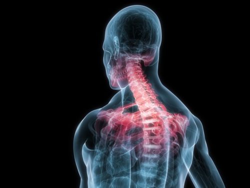
What is whiplash?

Whiplash


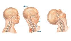
Whiplash is a quick, sudden hyperextension and then hyperflexion movement of the neck. The most common cause is a motor vehicle accident, although it also can occur in contact sports and falls.

The sudden movement causes the muscles, tendons and ligaments to be stretched beyond their normal limits, resulting in tissue damage and a generalised pain in the neck, upper back and even sometimes in the lower back.  The reason for this generalised pain is that the nervous system is in shock, due to the extreme sudden movement of the neck. Restricted movement results, which is the nervous system protecting the spine from further damage.


Whiplash associated disorder (WAD) is categorised into four stages:

  • WAD I – Neck pain with stiffness or tenderness, no physical signs
  • WAD II – Neck pain with musculoskeletal signs, such as decrease in range of motion
  • WAD III – Neck pain with neurological signs, such as numbness, pins & needles or weakness of the upper limbs
  • WAD IV – neck pain with a fracture or dislocation of the neck

The most common presentation to a chiropractor is WAD II.

There is also other symptoms involved with whiplash, which include, but not limited to – headache, arm weakness, jaw pain, dizziness, vertigo and pain when swallowing.


Do the symptoms occur straight away?

Whiplash symptoms do not always occur straight away. There can often be a delay of a few days in which the patient will start to report stiffness and a generalised pain around the upper back and neck.

Is an x-ray or MRI needed?

Imaging depends upon the symptoms. For a WAD I & II, imaging is not often required, as the injury is soft tissue. If a fracture is suspected, an X-ray will be needed. If there are neurological symptoms present, MRI is often needed to see and identify what is causing the symptoms.

Do I need to wear a cervical collar to immobilise?

No! A collar is not recommended, unless there is a fracture suspected or neurological signs present. The best thing for whiplash that is diagnosed as WAD I or II post injury is to encourage gentle movement.

How long does whiplash last for?

The duration of whiplash is quite variable and varies between person to person. Typically, recovery occurs withina few weeks, although it can last for up to 6 months and possibly longer. Receivingappropriate treatment and performing the provided exercises ensures the best outcome.

What is the treatment?

After a thorough history and examination, an individual treatment program will be provided with appropriate manual therapy and rehabilitation exercises to help improve joint range of motion, decrease pain and loosen muscles. Joint manipulation is often used, although not always straight after a whiplash injury.

If we deem that chiropractic treatment is not suitable for your injury, a referral to an appropriate health care provider will be given.


If you are suffering from whiplash, please contact us on at Mona Vale Chiropractic Centre on 99975773 to make an appointment, so we can help you begin your recovery from a whiplash injury.


Mitchell Roberts – Chiropractor

Just how heavy is your child’s school bag?

I often ask parents how heavy their children’s school bag is and the reply almost every time is ‘far too heavy’. A great new research paper has just come out titled “The weight of pupils’ schoolbags in early school age and its influence on body posture”. I will summarise points below and there is also a link to the paper (full text is available) at the bottom of this post.


The study looked at 168 children aged 7-9 over a period of 1 year and looked at school bag weight, length of straps and certain body posture measurements.

  • School bag weight ranged between 4.7-9.0kg, with an average weight of 6.3kg. The recommended weight for school bags for students aged between 7-9 should be between 3.1-5.9kg
  • More than 90% of the students improperly put on and took off their school bags. This involved a bending motion accompanied by rotation and a rapid straightening of the lumbar spine
  • 79.3% had the two shoulder straps adjusted at different lengths
  • School bags were most often placed too high, which forced children to lean forward and hence overload the spine


Some general tips for carrying backpacks:

  • Backpacks should ideally be no heavier than 10% of the students body weight
  • Use both shoulder straps, both of even length. Do not carry it over one shoulder
  • Use the waist straps
  • Heavy items should be placed at the bottom of the bag and close to the spine
  • Lift a backpack through squatting, backpack held close to the body

If you have any concerns about the effects of your child’s backpack on their spinal health, bring them in to see one of the chiropractors here at Mona Vale Chiropractic Centre.


The weight of pupils’ schoolbags in early school age and its influence on body posture. Brzek et al. BMC Musculoskeletal Disorders (2017) 18:117

https://bmcmusculoskeletdisord.biomedcentral.com/articles/10.1186/s12891-017-1462-z


Mitchell Roberts – Chiropractor